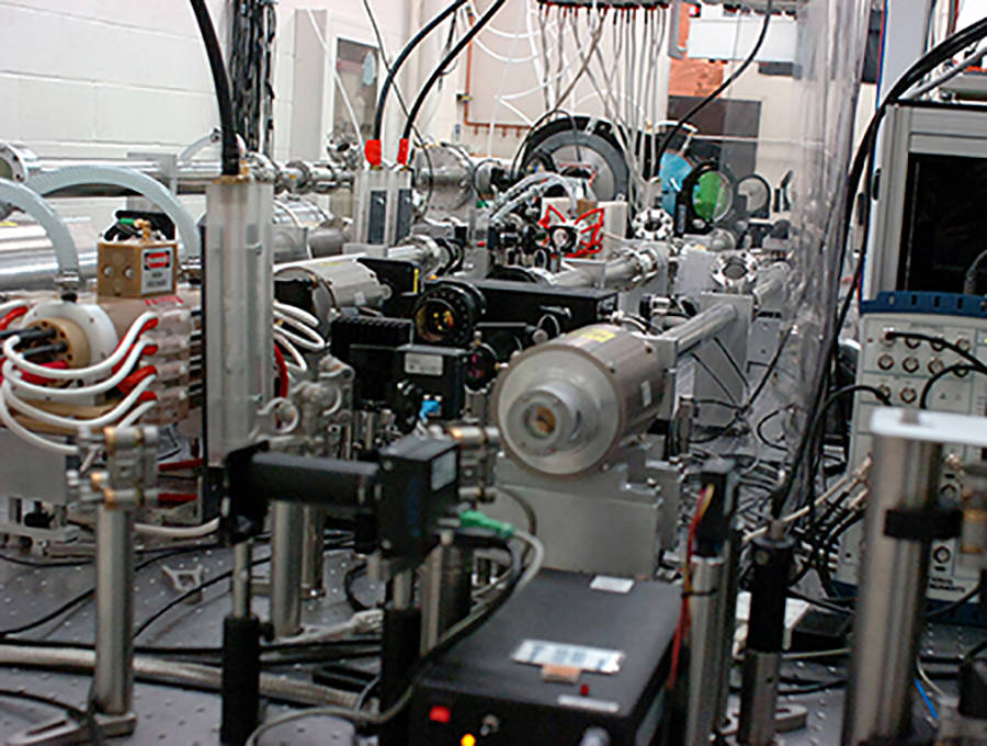
A team of scientists, led by University of Nevada, Reno’s Hiroshi Sawada, an associate professor of physics, demonstrated that numerical modeling accurately reproduces X-ray images using laser-produced X-rays. The images were obtained using the university’s chirped pulse amplification-based 50-terawatt Leopard laser at their Zebra Pulsed Power Lab.
The modeling approach established in this work could be used as a predictive tool to simulate radiographic images of complex 3D solid objects without carrying out radiation-based experiments.
The work illustrates a numerical method to model and predict X-ray images using widely available numerical tools.
A high-intensity laser can produce an intense X-ray beam in the laser-target interaction. Such laser-produced X-rays have been applied for recording X-ray images of various objects including a compressed laser fusion fuel, but a numerical tool for the quantitative comparison of a radiographic image has not been available until now.
“A challenge to a realistic simulation of laser-produced X-ray radiography is its spatial scale,” Sawada said. “Generally speaking, numerical modeling simulates physics phenomena in much smaller spatial scale than actual experiments. To overcome this limitation, we have separated modeling into two steps: X-ray generation is calculated with a fine resolution spatial grid, while computation of X-ray images using the calculated X-ray source is performed with a coarse grid to reproduce an X-ray image at a real experimental scale. Furthermore, a 3D computer-aided design-like model of a test object allows us to directly compare experimental and simulated images.”
Laser-produced X-ray sources could be an alternate source of non-destructive industrial imaging and medical imaging from soft tissues to heavy metal objects, the team of scientists have found through experiments with chirped pulse amplification using a tightly focused laser beam and several target materials.
The work, funded by the National Science Foundation, is published in Plasma Physics and Controlled Fusion. In this paper, the team presents experimental benchmarking of numerical modeling for fast electron and X-ray source characterization as well as broadband X-ray radiography. The work shows both qualitative and quantitative agreement between the experiment and the simulation for different X-ray attenuation filters.
In this paper, they present experimental benchmarking of numerical modeling for fast electron and X-ray source characterization as well as broadband Xx-ray radiography. The work shows both qualitative and quantitative agreement between the experiment and the simulation for different x-ray attenuation filters. The NSF-funded work is published in Plasma Physics and Controlled Fusion.
Sawada, a faculty member in the College of Science, and physics undergraduate student Chris Salinas started working on the modeling project in spring 2018.
“This work would have never been published without the help of students,” he said.
The team’s article, “Development of a predictive capability of short-pulse laser-driven broadband X-ray radiography,” was published by IOP Publishing in their journal Plasma Physics and Controlled Fusion in April.